Diagnosis of Newborn Brain Diseases Using Neurosonography
There are two ways to interpret the term.
- Neurosonography - most often this term means an examination of the brain of a newborn through the anterior (large) fontanelle.
- A group of methods for real-time research of the brain of newborns and adults, which also includes ultrasonography of the scalp, skull, spinal cord and brain, muscle ultrasonography and intraoperative ultrasonography. Ultrasound of the brain is very popular among practitioners.
In the future, transrodial ultrasonography will be considered.
Research principles
Since from the moment when the fontanelles close, it becomes impossible to carry out the NSH, it has diagnostic value only in children of the first year of life.
During the process of ossification of the skull, the large fontanelle closes by the age of 6 months - 1 year.
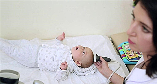
The study of the brain is carried out through the fontanelles: large (anterior), small (posterior), posterolateral and large occipital foramen. An ultrasonic sensor is installed at the site of the projection of these formations on the scalp.
The doctor, sequentially installing the sensor on these points, can examine all structures of the brain.
With the help of ultrasound, one can "see" the bones of the skull: ethmoid and nasal, orbital bones, assess the state of the interhemispheric fissure and spaces under the membranes of the brain, the structure and integrity of the brain substance, examine the state of the cerebral ventricles and measure the amount of cerebrospinal fluid in them.
The fontanelles in newborns are called "acoustic windows" in ultrasound diagnostics, because the cartilage by which they are formed, unlike the bone, is capable of transmitting ultrasound.
Informativeness
Only MRI can provide more accurate information about the state of the brain.
A large place among the causes of death in newborns is held by cerebral hemorrhages and ischemia. Transrodial neurosonography is used for these emergencies and is often the only method to confirm the diagnosis.
The size of the anterior fontanelle affects the ultrasonographic findings.
It is also difficult to diagnose meningeal hematomas, which pose a danger to a newborn and are one of the most common causes of emergencies.
Safety
The study does not require intervention in the baby's body, it is safe and painless. There are no reliable publications confirming the harm of ultrasound diagnostics for humans as a whole.
Who is NSG indicated for?

This is the main method for detecting structural pathology of the brain in newborns. The main indication for diagnostics is the deviation of the infant's neurological status, coupled with a violation of a number of laboratory parameters.
The method is invaluable if the child's condition is difficult and it is not possible to transport him to the functional diagnostics department for MRI.
Neurosurgical operations carry a high risk, since orientation in the cranial cavity and brain structures is difficult.
Ultrasonography can help the surgeon to ensure that the surgical instrument hits the target as quickly and accurately as possible, using a minimum of technical means.
Ultrasound helps to determine the localization, depth, size of the surgical target, to choose the optimal access with a minimal operating window.
Children of the first month of life
Performed if a pediatrician or neonatologist suspects or diagnoses the following conditions:
- intrauterine infection;
- prematurity;
- a history of hypoxia (during pregnancy or childbirth);
- anomalies in the development of organs or systems;
- anomalies in the development of the bones of the facial and cerebral parts of the skull;
- infectious diseases are found in the mother or child.
Children under one year old
In older children, the study helps to track the timeliness of brain development, to identify the cause of neurological symptoms (convulsions, pain, etc.).
It is worth remembering when carrying out an NSG that the older the child, the smaller the fontanel and narrower the acoustic window. Hence, a smaller viewing angle for the sensor.
For children under one year old, an ultrasonography method is available - visualization of the structures of the spinal cord. Spinal ultrasonography is used before lumbar puncture, as a screening method for diagnosing pathology of the spinal canal.

Is it performed by an adult?
Rarely, but fulfilled. In adults, the bones of the skull are connected by sutures, the fontanelles are closed, and there is no cartilage between the bones of the medullary skull. Ultrasound cannot penetrate into the cranial cavity, which makes research impossible (in the traditional sense, research through the fontanelles).
In adults, transcranial ultrasonography is possible. Diagnosis of cerebral edema, dislocation, and enlargement of the third ventricle is possible. But in some cases in adults, the brain is not available for research because of the narrow "acoustic window".
The use of the results of ultrasonography and the interpretation of the clinical picture makes it effective to diagnose intracranial hematomas, which are often present in adult patients with cerebrovascular accident.
But the main method of neuroimaging in adults and older children is CT and MRI.
How to prepare your baby for research?
The study does not require special preparation. Neurosonography is performed in the morning, the child is placed on a rigid couch, in a supine position. It is necessary to keep the child in a stationary position for some time so that the sensor does not "slide" from the area of the acoustic windows.
What are the contraindications for performing neurosonography?
There are no contraindications for the study. The procedure is painless, safe and has no contraindications on the part of the baby.
What happens during the research?
For research, an ultrasound source with a frequency of 5 MHz is used. For research through closed fontanelles or bones of the skull, lower frequencies are used, which affects the image quality and the information content of the study.
When examining premature infants, or to study the convolutions of the brain or intrathecal spaces, higher frequencies up to 10 MHz are used.
During the examination, the ultrasound emanating from the sensor is reflected from the media of the cranium, bones, and brain, which have different acoustic densities. These reflected waves are captured by the sensor, encoded and transmitted to the monitor screen as an image of intracranial structures.
The plane that the doctor can see depends on the method and location of the sensor.
From foci of compaction (tumor), softening of the brain substance (ischemia), areas of other tissue (blood), cavities in the brain substance (cysts), the reflected signal changes, which is recorded by the sensor. These changes are visible on the monitor screen.
What allows us to suspect or identify sonography in infants?
The doctor evaluates the following parameters:
- changes in the ventricles of the brain or their absence;
- evaluate the structure of the brain tissue (the presence of cavities, hemorrhages, tumors, ischemic foci);
- assess the state of blood vessels;
- assess the condition of the membranes and intrathecal spaces (expansion can speak of dropsy of the brain, for example).
Deciphering the results of cerebral neuropathy in newborns

The norm is not a specific figure, but the absence of some kind of organic pathology in an infant. Decryption is carried out only by a qualified diagnostician, neonatologist or pediatrician.
Normal values
For normal values of the size of the ventricles of the brain, values equal to twice the value of the standard deviation are taken. All parameters depend on how many weeks the child has developed in utero (premature or delayed), the norms are calculated based on this.
Also, the parameter depends on what day of the child's life the study is being carried out. Each clinical case has its own norm.
Deviations from the norm
The norm of indicators obtained in neurosonography cannot be considered a specific number. The peculiarity of rationing in children of any age is that there is an upper and lower limit of the norm, the values within which are not deviations.
All data obtained in the study of the brain can be assessed only by a neurologist, taking into account laboratory parameters and clinical picture.
Parents' tactics upon detection of pathology
The most important thing is not to panic, seek help from a specialist who will guide your child. Parents are required to follow all recommendations, monitor and inform the doctor about the change in the baby's condition.
Research cost
The cost of the study depends on the clinic in which it is carried out and the level of the specialist. On average, neurosonography can cost about 2,500 - 3,000 rubles.

Conclusion
Lesions of the central nervous system dominate among the general morbidity and mortality in newborns. Intraventricular hemorrhages are especially common. The shorter the period for which the child was born, the greater the risk of hemorrhage. LSS of the brain of newborns is a study aimed at early diagnosis of such pathological conditions that threaten the life and development of the baby.
The method itself does not pose a danger to the newborn and is not a cause for concern for parents.
Research is available as a paid service. It is carried out in the intensive care unit or neonatal pathology in the event of a number of emergency conditions, and ultrasound examination is also possible intraoperatively to study the brain in real time.
When performing a series of studies, it is possible to assess the dynamics of the pathological process (decrease in the volume of the hematoma or the size of the third ventricle after the interventions).