Hydronephrosis is understood as a persistent progressive increase in the size of the renal pelvis, calyces against the background of impaired urine outflow. Hydronephrosis in newborns is dangerous because it leads to gradual atrophy of the renal tissue and the development of renal failure. Treatment of this disease is surgical.

Hydronephrosis of the kidneys in babies
Hydronephrosis occurs in about 10% of infants. According to medical statistics, the disease occurs in boys several times more often. Due to hydronephrosis, the size of the kidneys gradually increases. Along with this, there is a noticeable thinning of the parenchyma (the main tissue of the kidneys, in which urine is formed). As hydronephrosis develops, the kidneys stop working.
Note! The most dangerous for babies is bilateral hydronephrosis. This severe defeat can provoke a fatal outcome. It occurs as a result of the accumulation of toxic metabolic products in the tissues of the body.
Causes of the disease
This disease can be congenital and acquired. Hydronephrosis of the kidneys in newborns from birth occurs for the following reasons:
- abnormal location of the renal arteries;
- compression of the ureters;
- impaired motility of the urinary tract;
- abnormal location of the ureter (when it is localized behind the vena cava);
- congenital blockage of the urinary tract.
Common causes of acquired hydronephrosis are urolithiasis, pyelonephritis.
Disease development
The development of hydronephrosis occurs gradually. Parents note the presence of its signs when the child's renal parenchyma is already affected.
When develops
With an incorrect structure of the urinary tract, the disease begins to develop from the first weeks of a baby's life. When there are inflammatory pathologies of the urinary tract, then the first signs of a possible progression of hydronephrosis can be seen in 1-2 years.
What causes the pathology
A very common cause of the appearance of pathology lies in urinary reflux. In this case, urine is thrown back into the ureter. The baby has difficulty urinating.
Hydronephrosis also often appears due to impaired conduction of nerve impulses. The disease sometimes occurs due to improper formation of the nervous system during intrauterine development.
Some babies have a megaureter, which is an enlarged ureter. This feature of its structure adversely affects the processes of urine separation.
Signs
Hydronephrosis is characterized by the following pronounced symptoms. They increase as the intensity of the painful process increases:
- Violation of urine separation. Due to the destruction of the kidney tissue, its amount decreases.
- Soreness in the abdomen.
- When probing, a volumetric formation is noted in the hypochondrium.
- The appearance of blood impurities in the urine.

Blood in the urine of a newborn
- Increase in body temperature. It rises significantly when an infectious process is attached.
All these symptoms are combined with edema. The accumulation of fluid in the body adversely affects its performance.
Pathology stages
Experts distinguish 3 stages of hydronephrosis development:
- In the first stage, kidney function is practically not affected. Laboratory data reveal minor changes in the work of the excretory system. Parents may not suspect that pathological processes are taking place in the baby's body.
- At the second stage, a slight increase in the size of the renal pelvis is found. In parallel with this, there is a thinning of the renal tissue. Renal urine production is reduced by about 40%.
Important! At this stage, the kidneys can still be restored. However, in the absence of the necessary treatment, organ performance will rapidly deteriorate.
- At the third stage of development of hydronephrosis, the kidney increases by about 2 times (relative to the norm). Its functionality is reduced by 4/5.
In terms of severity, this pathology is divided into four degrees:
- In first-degree hydronephrosis, the kidney tissue is not damaged. A qualified specialist can detect this disease.
- With hydronephrosis of the 2nd degree, the parenchyma changes to an insignificant degree, however, laboratory test data show characteristic changes in the blood picture.
- With grade 3 disease, there is a strong lesion of the renal parenchyma. It is at this time that pronounced clinical symptoms occur.

Changes in the kidney with hydronephrosis
- With grade 4 hydronephrosis, the kidney tissue is almost completely destroyed.
Diagnostics
Most often, newborns have left-sided hydronephrosis. This is due to the fact that, due to some anatomical features of the structure of the excretory system, the left kidney is slightly higher than the right one.
Diagnostics includes the following measures:
- Analysis of urine.
- X-ray of the lumbar region (using a contrast agent).
- Ultrasound examination of the kidneys (it allows you to determine the size of the organ and the state of its parenchyma).

Ultrasound of the kidneys for a newborn
- Computed tomography (it allows you to make an accurate diagnosis in doubtful cases).
If these types of examinations do not make it possible to make an accurate diagnosis, then the child is sent for urography. This is a rather risky measure, because the contrast agent used in this type of diagnosis can poison the baby's body.
Treatment
Hydronephrosis can only be cured with surgery. Moreover, the earlier a child was diagnosed with hydronephrosis, the more chances of a successful outcome. Treatment with folk remedies can only be used as an auxiliary method.
Attention! Ignoring surgical treatment and replacing it with drugs of "traditional medicine" lead to the loss of precious time.
Operational activities
Surgical intervention is aimed at correcting the diameter of the ureter. Stenting is often used for this. With a megaureter, an operative narrowing of the ureter occurs.
With grade 3 or 4 hydronephrosis, urgent surgical intervention is required. Moreover, it is performed even in cases where only one kidney is affected by the disease. The criterion for a successful operation is the restoration of urine outflow.
The main types of surgical interventions in the treatment of hydronephrosis are as follows:
- Pyeloplasty. During the operation, the affected tissue is first removed, the narrowed areas of the ureters. Then the ureter and pelvis are artificially connected.
- Antevasal pyeloplasty is performed in the presence of an additional vessel. In this case, a message is formed on top of it between the ureter and the renal pelvis.
- Laparoscopic surgery is characterized by minimal trauma. The kidney is accessed only by small punctures in the abdominal wall.

Kidney surgery in a newborn
Drugs
Medicines for hydronephrosis are not prescribed. This is due to the specificity of the disease. It is possible to cure diseased kidneys only with the help of surgical intervention.
Can I pass by myself
This disease cannot go away on its own without appropriate treatment. In the absence of treatment, the following complications may develop:
- pyelonephritis;
- acute renal failure;
- atrophy of the renal parenchyma.
Important! With atrophy of the renal parenchyma, even surgery will not be able to normalize the normal outflow of urine. The child will need long-term treatment.
Postoperative rehabilitation
With a successful operation, the recovery period does not last long. It takes only two weeks for this. After that, the child remains for some time under the supervision of a pediatrician and urologist. Postoperative rehabilitation may take longer if there are complications.
Prevention
The development of hydronephrosis can be prevented by:
- regular visits to the ultrasound office during pregnancy;
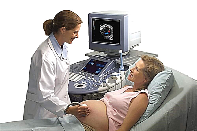
Ultrasound of a pregnant woman
- observation of the newborn by a neonatologist;
- timely access to a doctor if there are signs of pathology.
No other specific prevention methods have been developed.
Hydronephrosis of the kidney in an infant is a serious illness. It has no other methods of treatment, except for surgery. Parents should carefully monitor the condition of the baby. When the first signs of kidney disease appear, you should immediately contact your pediatrician or urologist.