A computed tomography (CT) scan of the brain may be required for a child at any time. Not only suspicions of diseases of the brain and nervous system, but also head injuries of various origins can force the doctor to issue such a referral. In this article we will tell you how a CT scan is performed and what it shows, and also tell you how to properly prepare a child of any age for examination.
What it is?
With the advent of computed tomography, physicians were able to study any internal organs of a person without injuring him, without violating their integrity. CT makes it possible to study a specific organ in layers and in detail. The essence of the method is based on the fact that tissues of different density reflect X-rays in different ways. This allows you to get a very detailed picture of the organ and all its inner layers and detect even the smallest deviations in its condition. For the development of the method in 1972, scientists received the Nobel Prize.

The technique differs from MRI in the type of rays. In MRI, the study is carried out using electromagnetic radiation, in CT, X-rays are used. It would seem that there is less harm from electromagnetic radiation, why not examine children for MRI? The answer is simple: MRI is not always accurate and can detect a problem. CT accuracy is higher.
Even though the radiation doses for diagnosis in children are minimal, the examination is done strictly according to indications and only when the harm from the diagnosis itself does not exceed the harm from its absence. There is some harm from X-rays, but severe pathologies of the brain, if suspected of which a CT scan is prescribed, can harm a child much more than a single exposure to minimal doses.

How old is it?
An examination can be prescribed for a child of any age - a newborn, an infant, a child who is already 3 years of age or older. The baby is more likely to undergo neurosonography (ultrasound of the brain). And only if preliminary studies show deviations, confirmation or refutation of the presumptive diagnosis by CT may be required.
The peculiarity of computed tomography of the brain in early childhood is that it uses means to immerse the child in deep drug sleep (anesthesia). During the examination, the patient must lie completely still for quite a long time, and there is no practical possibility to force the baby to do this.

Indications for use
As already mentioned, computed tomography can be prescribed only in extreme cases. They try not to use this method unnecessarily. Such good reasons are considered:
- severe birth trauma to the head (depressed fracture of the skull, displacement of bones, injury or extensive hematoma of the brain with hemorrhage, etc.) - the study is carried out in the very first hours after the birth of the child;
- injuries from falling on the head (if there is a suspicion of a pinched brain, injury to its individual parts, fractures of the skull bones) - the study is carried out as soon as possible after hospitalization due to injury;
- prolonged intracranial hypertension of unexplained origin (allows to establish the reasons) - is carried out after the condition has been diagnosed by other methods several times in a row;
- mental disorders and diseases (to confirm the diagnosis and search for the cause) - carried out in the direction of a psychiatrist for children over 3 years old;
- tumors, cysts, neoplasms any genesis in the region of the brain - a study is carried out after confirming the presence of a tumor by other methods to establish the nature and extent of education;
- vascular disorders, acute and severe cerebrovascular accident;
- paralysis, paresis, impaired motor functions (both congenital and sudden onset).


Contraindications
A CT scan of the head of a child will not be performed if he is allergic to substances used as contrast agents, such as iodine. Children with severe renal failure are also not examined.
If there are metal objects in the body, which can be implants, staples, CT is also not performed. The diagnosis may also be refused if the child has a severe mental disorder, accompanied by an inadequate response to drugs for anesthesia. Contraindications include severe forms of diabetes mellitus, myeloma of the skin and some pathologies of the thyroid gland.


How is it going?
To carry out such diagnostics, medical institutions are equipped with special rooms equipped with a modern tomograph. It is a very sophisticated device, thought out to the smallest detail, equipped with sensors with incredibly high sensitivity to reflection of X-rays. The data from the detectors are sent to the computer, where the image is formed. Very complex and extensive computer programs help the doctor to correctly and accurately decipher the data obtained.
The child is weighed so that the anesthesiologist can accurately calculate the amount of the drug to be injected for immersion in a deep medication sleep. Then a contrast agent based on iodine is injected into the baby's vein. When examining the brain, diagnostics with contrast is considered the only accurate one.
If you can get the results of CT scan of bone tissue, for example, of the spine, without a contrast agent, then a study of the brain without contrast will be uninformative. Older children are not given anesthesia.

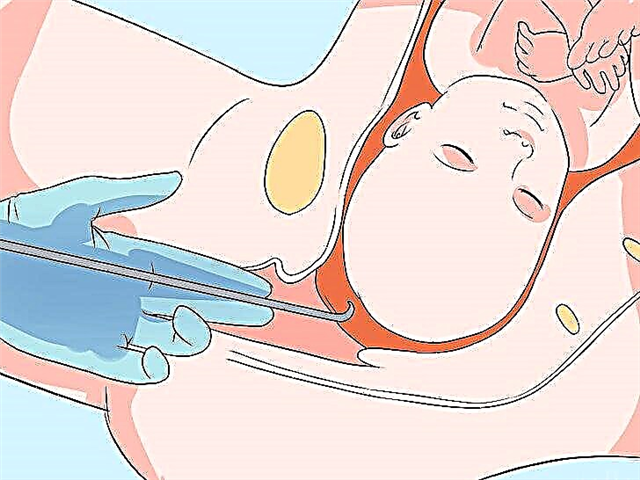
The child is placed on the tomograph table in a supine position. The head is fixed in the required position with soft straps. The table is pushed into the tomograph capsule. Mom and other people are asked to leave the office while the hardware complex is operating.
The scan lasts from 30 minutes to one and a half hours, the results will be ready in about an hour after the end of the examination, in emergency cases - in 20-30 minutes.

Do you need preparation?
Preparation is required without fail. For 3-4 hours before the examination, the child is not given food and water if immersion in medication is to be expected. A child who, due to his age, does not need anesthesia, can be fed about a couple of hours before the CT scan.
A child who is to undergo the procedure without anesthesia must be explained that nothing terrible and painful will happen. Chad can be invited to play in a spaceship, because the tomograph really resembles the scenery from "Star Wars". Be sure to warn that the main rule is the absence of movements and movements during the examination.

In order for the child to breathe fully during the study, vasoconstrictor drops must first be dripped into the nose. Provide the doctor with all the required tests before the diagnosis: a pediatrician's conclusion, ECG results, a general blood test and the same urine test.
If a child is allergic or is very young, so that doctors have enough information about the allergic status, it is recommended to visit an allergist or pediatrician and make tests for iodine-based contrast agents. A certificate of the absence of a negative reaction to iodine is also provided before starting the study.
If CT is done on an emergency basis, for example, due to an injury, preliminary preparation is omitted, limited to only a short rapid test for allergy to a contrast agent.

Reviews
According to mothers, the procedure scares not only children, but also parents. Mothers fear for the consequences of X-ray radiation, as well as for finding the child in the tomograph capsule if he is not given anesthesia due to his age. It happens that a 10-12 year old child makes a real scandal, refusing to go to the capsule.
In many children's hospitals in Russia, open tomographs are being installed for children, in which the child should not be in a confined space. Such a tomograph shows everything necessary just as well, but psychologically children perceive it better and easier.
Children, according to mothers, rarely tolerate anesthesia easily. Waking up is often unpleasant.
You will learn more about how a CT scan of the child's brain is performed in the following video.


