The brain is considered one of the most complex and mysterious human organs. The quality of life and developmental characteristics of the child depend on his condition and health. Therefore, a special control is established for the brain, which begins from the very birth of a child. The neurosonography method helps to find out whether the baby's brain is formed correctly and is developing normally. We will tell you about how the survey is carried out and what it shows in this article.
What it is?
Neurosonography is an ultrasound scan of the brain of babies. In fact, this is an ordinary ultrasound scan, well known to everyone, but it is only performed at the most convenient period, when the baby's “fontanelles” have not yet closed.

The mobility of the bones of the skull is necessary for the baby to ensure the passage of the head through the birth canal of the mother during birth. And for quite a long time, the “fontanelles” remain open. It is this feature that makes it possible to conduct an ultrasound scan of brain structures in search of signs of congenital or acquired ailments.
Usually, cerebral NSH is performed in newborns and children under the age of one and a half years, after this age, the "fontanelles", as a rule, close. After that, for some time, it is possible to study through the temporal lobes, and then it will be possible to obtain information about the state of the brain only through electroencephalography (EEG), computed tomography (CT) or MRI.

Neurosonography has been included by the Russian Ministry of Health in the list of complex ultrasound examinations of the first screening, which is performed on infants at 1 month old. Earlier and later than this period, the examination is carried out in the presence of medical indications, which we will discuss below.
Is it safe?
Despite a lot of rumors, most of which are generated by parents themselves in numerous forums on the Internet, the study is considered safe and harmless for babies. The essence of what is happening is quite simple, and to understand it does not require deep knowledge in the field of physics: the sensor generates ultrasonic waves of a certain frequency and length, the waves pass perfectly through the tissues of the human body, are reflected from different brain structures in different ways and, reflected, are sent back ...

The sensor catches the "response" and forms an image on the monitor of the ultrasound machine. It is this image that the doctor assesses, but again not "by eye", but using special mathematical algorithms inscribed in the scanner software.
Rumors about harm, it is possible, were not born out of nothing, because medicine that has been using the ultrasound method for only about 20 years does not yet have a sufficient statistical base to prove that there is no harm from ultrasound exposure in the future. Collecting such information takes well over two decades.
However, there are also no data confirming the harmful effects of ultrasound studies on the child's body. Therefore, the procedure is considered safe. She doesn't hurt the baby. Opponents of the study should be reminded about ultrasound during pregnancy, because part of the fetal brain structures were assessed through the anterior abdominal wall of a pregnant woman. But neurosonography of the infant allows you to get a more complete picture of the structures and functioning of his brain.

If necessary, the baby can do neurosonography as many times as it takes to establish or clarify the diagnosis until the fontanelle heals and the bones of the skull begin to become strong.
Technique
Technically, neurosonography is not much different from any other ultrasound examination. The child is placed on a diaper, laid on a couch in a supine position. The doctor applies a small amount of acoustic gel to the fontanel area for a tighter fit of the transducer and better conduction of ultrasonic waves. Within 7-10 minutes, the sensor moves over the baby's head, measurements of individual parts of the brain are made, after which the parents are given the NSG protocol.

There is a NSG with extended capabilities - with a Doppler. This study gives an idea not only about the structures, shapes and sizes of areas and parts of the brain, but also about the process of blood supply to this most important organ.
Preparation for neurosonography is not required. The only limitation concerns anti-spasmolytic medications and analgesics. Such funds should not be given to the child a couple of days before the examination, since they affect the size of the vessels.
Mom may well feed the child before the examination, so that the baby does not worry and let the doctor examine himself. But even if your little one bursts into tears during the scan, this will not affect the results in any way: neither the size, nor the functions of the brain regions change from the baby's behavior.

Who needs it?
As already mentioned, it is advisable to do an NSG for all babies at 1 month or 3 months, if for some reason a medical examination of the child at four weeks of age was not carried out.
The advice of the Ministry of Health is, of course, advisory in nature, and therefore parents can refuse to be examined, but it is not recommended to do this, because in the presence of pathologies later, when the "fontanelle" begins to close, the diagnosis will be difficult.
However, there are categories of babies for whom neurosonography is especially recommended. First of all, these are children who were born prematurely (up to 37 weeks of pregnancy inclusive). Premature babies are a category of special risk, including the likelihood of developing pathologies from the brain and central nervous system. Experts also consider it compulsory to carry out NSG for children whose appearance occurred by surgery - if a woman had a cesarean section.


If the following symptoms are found in a child during the first weeks of life, parents should also not give up neurosonography:
- the child behaves strangely - in the absence of diseases, he eats poorly, often spits up profusely, he is inactive, does not show vivid emotions, often cries, sleeps superficially, constantly wakes up, if the baby has a pronounced tremor of the limbs, chin, there is a squint;
- the baby often cries, throws his head back and bends his back (this may be an indirect sign of increased intracranial pressure);
- the baby does not hear well or reacts poorly to visual stimuli, does not follow the toy with his eyes, cannot focus his gaze on the mother's face;
- low blood pressure in a baby, fainting, seizures;
- severe coordination disorders (infant flapping and flinching has nothing to do with it);
- the child has a birth trauma, or he fell, hit his head, there were sharp throwing back of the head after birth.
If the child is scheduled to undergo an operation on the vessels or heart in the near future, the NSH is done without fail. An unscheduled neurosonographic study will be carried out in the event of a fall, because the method allows you to establish signs of a concussion, bruise, or the fact of the formation of cerebral hematomas.
A child who has had a severe viral infection should also be examined to rule out signs of encephalitis or meningitis. The NSG method is also used in the diagnosis of tumors.
It is imperative to examine babies who were born with a low weight (less than 2700 g), as well as babies who were born with asymmetry (in which one ear is lower than the other, one eye is larger than the other, etc.)
External anatomical malformations (the presence of extra fingers and toes, the absence of limbs, etc.) are also a good reason for a careful ultrasound examination of the baby's brain.
Children who were born after pregnancy, which was accompanied by fetal hypoxia, Rh-conflict, must undergo an NSG, since the long-term consequences of these unfavorable intrauterine conditions can be quite severe.
Indicators of the norm
In a healthy child under one year old, and in older children, both hemispheres are symmetrical. It is this indicator that the doctor evaluates and describes first. Symmetry violation can be a sign of both congenital anomalies and the development of a tumor process.

In a healthy child, the grooves and convolutions of the medullary cortical layer are well visualized, without exception, all structural units are distinguished by clear and even contours. No fluid is found in the spaces between the right and left hemispheres in a healthy toddler.
The ventricles, the cistern have certain dimensions that correspond to the tables according to which the indicators are deciphered. Radiant beams show signs of hyperechoicity.

The ventricles of the brain, as indicated in the study protocol of a healthy baby, have a homogeneous structure, without foreign inclusions. If mom and dad are very interested in the meaning of the numbers in the conclusion, then the normal indicators are as follows:
- lateral ventricles - anterior horns - 2 mm (after 3 months - 2-4 mm);
- lateral ventricles-posterior (occipital) horns - 10-15 mm;
- the body of the lateral ventricles - no more than 4 mm;
- the size of the third ventricle of the brain is 3-5 mm;
- fourth ventricle - no more than 4 mm;
- interhemispheric gap - 3-4 mm;
- cisterna magna - maximum 10 mm;
- subarachnoid space - an average of 3 mm.
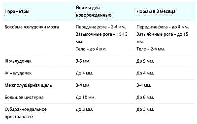
These data are not the ultimate truth. When a medical conclusion is made, the doctor must take into account the height and weight of the child, because in a small child with miniature shapes and sizes of the brain regions may differ downward.
Pathology
By refusing to be examined, parents run the risk of not seeing it on time, and, therefore, not starting timely treatment in the event of cyst-type neoplasms. Cysts can be different - some, for example, arachnoid, are quite dangerous for the baby and definitely need treatment.
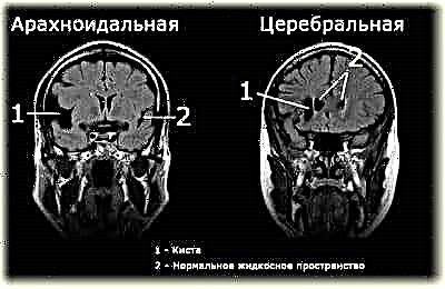
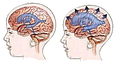
An increased volume of cerebral fluid inside the skull may indicate the presence of dropsy of the brain, and darkening and vascular pathologies may indicate ischemia, cerebral palsy, hematomas. The appearance of such terms in the ultrasound protocol is not yet a diagnosis, since additional diagnostics are required to make a diagnosis, neurosonography alone is not enough.
Quite often, such an examination reveals physiological and functional disorders that do not need any therapy and disappear over time on their own. They are due to the neurophysiological immaturity of the brain - a condition that is quite natural for newborns.
Some formations have a favorable prognosis, but need regular monitoring, and some conditions, such as hydrocephalus, need therapy as soon as possible.
Do not assume that a child who visually gives the impression of being completely healthy cannot have abnormal brain development. They are quite insidious and it is almost impossible to see them with the naked eye, unless, of course, the pathologies are of a total nature. We say this not in order to scare the parents, but in order for them to think well before giving up neurosonography, considering it unnecessary and even harmful.
Mom's opinions
Having received a referral to NSG, many mothers rush to the Internet for an answer to the question whether such a diagnosis is needed at all. And then they may well stumble upon reviews of a pseudo-scientific sense, in which opponents of ultrasound for a child justify the harm and destructive consequences of neurosonography for the baby. If you have a great desire, you can read such reviews, but we strongly do not recommend taking them on faith.
It is best to ask your doctor about the appropriateness and possible dangers of diagnostics.

There is a category of mothers who do not trust doctors from the district children's polyclinic. For them, there is also a way out - to do an NSG in a private clinic, however, already at their own expense - on average in the country such an ultrasound scan costs from 1,500 to 3,000 thousand rubles.
Moms who describe the "incredible suffering" of a baby during neurosonography do not take into account that the child is crying in the diagnostic room not from pain, but from fright, because a completely stranger is touching him, which, from the point of view of the baby, is a direct threat to him security.
For more information on the procedure for neurosonography in newborns and infants, see the following video.