
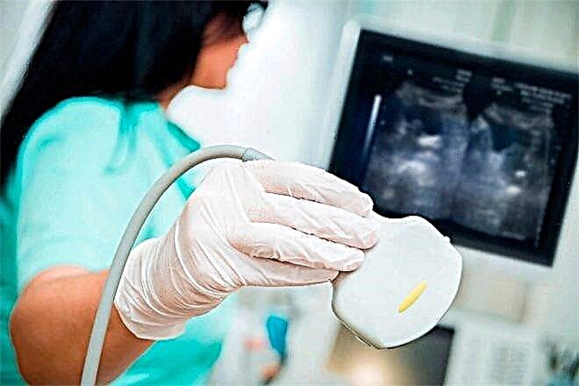
With the help of ultrasound, pregnancy can be determined quite early. Many mothers-to-be have many questions about how such research is carried out and whether it could be dangerous for their baby. This article will help you figure it out.

Pros and cons of research
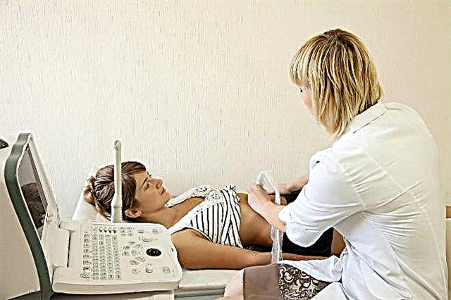
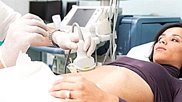
Currently, there are a wide variety of ultrasound methods that allow you to establish pregnancy even at the earliest stages. Screening is indicated for all women who have a suspicion that they will soon become mothers. This research is extremely important and necessary.
Ultrasound diagnostics is basic in establishing pregnancy. Carrying out it at certain stages of fetal growth is mandatory. This allows you to monitor the dynamics of its intrauterine development and identify various anomalies, as well as deviations at the earliest stages.

However, there are also disadvantages to this procedure. Of course, the human factor belongs to them.
European doctors have found that the discrepancy in the assessment of the results obtained can reach 20%. This is a fairly high rate, especially when it comes to pregnant women and their unborn babies.
There is also a risk of infection of the baby during an ultrasound scan through the vagina. It should be noted right away that this situation occurs extremely rarely and completely depends on the competence of the doctor conducting this study. If the doctor has the proper experience and education, then this situation is almost impossible.

Expectant mothers should remember that ultrasound is one of several diagnostic methods and is performed by a person. This implies that the results obtained are not 100% reliable. In some cases, they do not completely coincide with the real health indicators of the expectant mother and baby. In this case, it is required obligatory rechecking and conducting research with another specialist.
Kinds
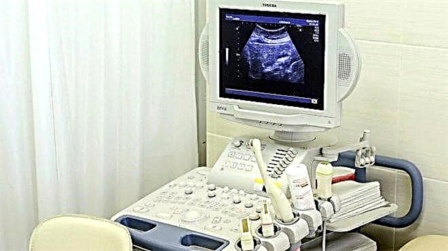
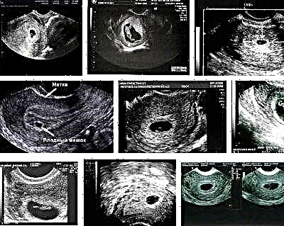
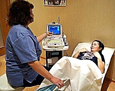
Early ultrasound techniques can be very different. The choice of research largely depends on the level of the material and technical base of the medical institution. It must be said that lately even the most ordinary district gynecological polyclinics have been equipped with rather modern devices.
Many expectant mothers do not know which method is better to detect pregnancy in the early stages. This choice is individual and depends on each specific situation. Usually, the first ultrasound technique is mandatory agreed with the obstetrician-gynecologist, which will guide the woman during the entire period of her pregnancy.

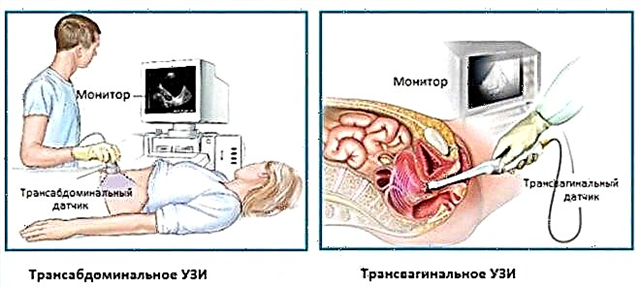
Surveys can be carried out using various types of sensors. Doctors call a vaginal probe examination transvaginal Ultrasound. You can also conduct research through the stomach. This method is called transabdominal.
The need for an ultrasound of the uterus or small pelvis is determined individually by the obstetrician-gynecologist. For this, all pathologies of the genital organs of a pregnant woman are assessed. The doctor who will observe the future mother in the future draws up the necessary diagnostic scheme for her during this period. As a rule, in most cases, combined research methods are used.
What indicators are being evaluated?
Expectant mothers should understand several basic concepts used by both ultrasound diagnostics doctors and obstetricians-gynecologists. They often use the term "Obstetric gestational age"... This concept implies the term for the development of the fetus. It is always calculated in weeks and days, not monthly.
Many doctors of ultrasound diagnostics use the term "embryonic term", which significantly confuses the expectant mother. It should be remembered that only the obstetric method of calculation is used to estimate the gestational age. Modern ultrasound machines automatically calculate it according to the basic parameters that are entered before carrying out this research procedure. Further the obstetric term is also used to assess the course of pregnancy.
Ultrasound examination in the earliest period of intrauterine development is done for:
- the establishment of a gestational egg in the uterine cavity, which means pregnancy;
- determining the stage of development of the embryo during its development;
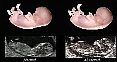
- identifying specific signs of a "frozen" pregnancy;
- establishing various violations and intrauterine anomalies.

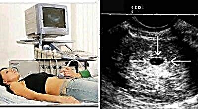
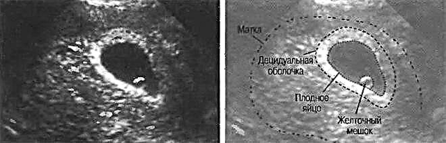
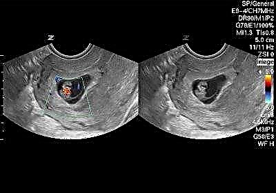
About the gestational egg
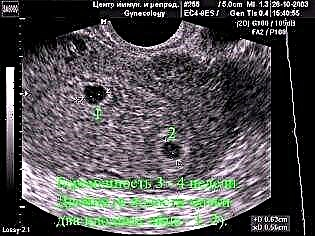
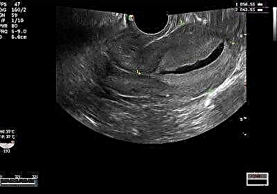
It is also called fertile. This is a characteristic criterion indicating that a woman is pregnant. Most often, it can only be detected by five weeks of intrauterine development. Some qualified and experienced professionals can detect the presence of a gestational egg in the uterus as early as 3 weeks.
Usually during this period you can set gestational age with an error of about 1 week. It is extremely difficult to identify any deviations in development at this stage. The first ultrasound will only show pregnancy, but will not be able to identify all the developmental anomalies in the fetus. Their doctors determine a little later - in the 2nd and 3rd trimester of bearing a baby.
Experts assess several basic parameters identified at the earliest stages of gestation.
They allow doctors to understand if the intrauterine development of the fetus is normal. The development of the embryo can be determined by determining its diameter. For this, as a rule, only one measurement is sufficient.
The average diameter allows you to determine the size of the gestational egg more accurately. This requires at least three measurements. Many mothers are interested in why it is also impossible to measure only one parameter. Such a study will not be informative and will not allow obtaining an accurate result.
If the gestational egg is determined at 4 weeks and three days after the first day of the last menstruation, then its size is usually 2-3 mm. At 5-6 weeks of intrauterine development from a similar day of calculation, the diameter already increases to 0.5 cm.Thus, the definition of this parameter is quite informative and allows you to track the dynamics of fetal growth.
These indicators will also help expectant mothers to calculate the approximate menstrual period of pregnancy. Usually, doctors call this term the obstetric period, but in the first weeks of bearing an unborn child. In this case, to determine the menstrual age, add 30 to the average diameter of the ovum (in mm). If this average diameter is greater than 16 mm, then 35 is added to the value.
The growth of the gestational egg in the first trimester is quite rapid. This feature is due to nature. It is at the earliest stages in the unborn baby that all vital organs are laid. This time is very important for every child.
The gestational egg grows at a rate of 1.8-2 mm every two days from 4 to 9 weeks of intrauterine development. It should be noted that this indicator for assessing the development of the future baby is not assessed, but is informative.
Doctors identify several clinical situations that should alert expectant mothers. If, with a size of 15 to 25 mm, the gestational egg in the uterine cavity is not detected, then this may be a sign of a "frozen" development of pregnancy. This symptom is extremely unfavorable. If this situation has occurred, then a pregnant woman, first of all, should not panic. In this case, it is required compulsory ultrasound control after 7 days.
If the size of the ovum is too large for a certain period, then this is also an extremely unfavorable symptom. Doctors believe that this may be a manifestation of the pathological course of pregnancy. This condition occurs when Frozen pregnancy or at empty egg syndrome... Only obstetricians-gynecologists detect these pathologies. In this case, it is categorically impossible to rely on only one ultrasound result.
The size of the ovum should moderately increase over time. If the opposite process is observed, then this may be an indirect sign of low water. It should be noted that the amount of amniotic fluid using ultrasound is determined much later. Usually, such a study is carried out only at 18-20 weeks of intrauterine development of the fetus.
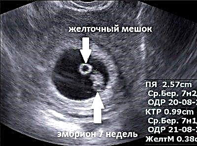
About the yolk sac
This anatomical formation appears even before the full formation of the embryo. Doctors consider the appearance of this clinical sign to be a reliable confirmation of the presence of uterine pregnancy in the female body. Some unqualified specialists of ultrasound diagnostics at this stage may be mistaken and not "see" an ectopic pregnancy.
The yolk sac is located between the chorion and the amnion. In the future, the placenta and fetal membranes will develop from these anatomical structures. The specific place in which the yolk sac is located is called chorionic space.
The size of this formation is associated with the parameters of the gestational egg. If the ovum is 0.5 cm in size, then the yolk sac may be about 6 mm. A variant of the norm can be considered a size from 3 to 5 mm.
The largest size of the yolk sac is at 10 weeks of intrauterine development. By this period, it grows to 0.5 cm. In the future, this formation also participates in organogenesis - the intestines of the unborn child are formed from it.
About amnion
Doctors consider this formation to be a special membrane (shell), which is located in the ovum. As a rule, this anatomical formation is clearly visible up to 11-12 weeks of intrauterine development of the fetus. During this period of pregnancy, the size of the fetus is about 5-7 mm. The complete completion of the formation of the membranes occurs only by the end of the 16th week of intrauterine development.

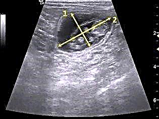
In addition to the yolk sac, amnion and ovum, ultrasound doctors determine a number of other important indicators. One of these parameters is determination of the coccygeal-parietal size. This indicator is described in the conclusion using a few letters. It can be called KTP or CRL.
The KTP parameter allows you to determine embryo length. It should be noted that when determining this indicator, ultrasound specialists quite often make various mistakes. In some cases, technical errors of the devices can also lead to an incorrect result. It should be noted that this occurs in cases where outdated equipment is used for ultrasound diagnostics or an inexperienced doctor is conducting the study.
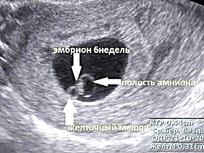
Using a correctly defined coccygeal-parietal size, it is possible to determine exact gestational age... The accuracy of the determination in this case can be even 3-5 days. If the size of the ovum is already 0.5-1 cm, then the immediate size of the embryo can also be determined, which becomes equal to 1-2 mm. In the future, every day the future man grows at a speed of about 1 mm.
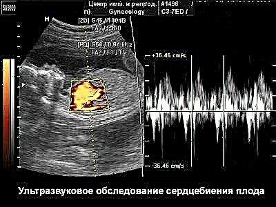
About heartbeat
Fetal heartbeat is another characteristic criterion that is determined in early pregnancy. This indicator is extremely important. Fetal circulation helps to assess its growth and development. It is possible to determine the heartbeat in the fetus already at a gestational age of 6 weeks.

Sometimes this indicator may not be determined. You shouldn't panic in this case either. In such a situation, a second ultrasound scan is required. It is usually carried out after 4-6 days.
The heart rate increases as the embryo grows. Up to 6 weeks of intrauterine development, this figure is usually 100-116 beats per minute. By week 9, the heart rate rises to 145-160 beats per minute. After 9 weeks, this figure begins to decline slightly.
A decrease in heart rate in the early stages of intrauterine development is usually an unfavorable indicator. Doctors call this condition bradycardia... The appearance of this symptom may indicate a pathological course of pregnancy and even its "fading". Any decrease in heart rate requires urgent intervention by gynecologists.
In the early stages of pregnancy, bradycardia can be determined by several criteria:
- if the coccygeal-parietal size is less than 0.5 cm, and the heart rate is less than 80 beats per minute;
- if the coccygeal-parietal size is from 0.5 cm to 9 mm, and the heart rate is less than 100 beats per minute;
- if the coccygeal-parietal size is 1-1.5 cm, and the heart rate does not exceed 110 beats per minute.
About the collar area
Collar size is another indicator that is used to determine the size of the embryo. Such an anatomical formation is a collection of lymph located between the skin and soft tissues of the embryo. The normal parameters of this zone are an important criterion for assessing various chromosomal pathologies that can develop in the fetus.
The determination of this indicator is carried out, as a rule, at 11-14 weeks. This test is part of genetic screening. Also, for additional diagnostics, a number of biochemical studies are carried out. This helps to establish the presence of any genetic abnormalities in the female body.
It is very important to conduct research during a certain period of pregnancy. Only a timely assessment of the results allows us to assess the real state of the fetus in the womb. At a later date, a different indicator is used. It is called the neck roll.
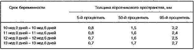
The measurement of the thickness of the collar zone is compared with the coccygeal-parietal size, equal to 45-84 mm. Compliance with the time criteria is very important and is due to the physiological development of the lymphatic system. The metabolism in the lymph is very fast. Normally, the thickness of the collar zone in this period of pregnancy is 3 mm. The pathological value can be considered a size of 0.5 cm at 16-18 weeks and more than 6 mm at 19-24 weeks.
About the nasal bone
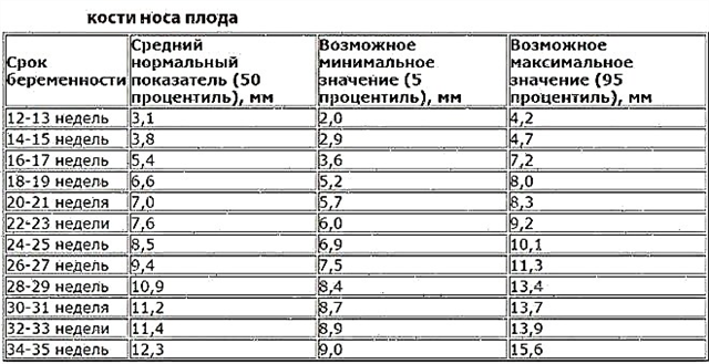
The nasal bone is another indicator that doctors evaluate in the very early stages of pregnancy. Such a study helps to identify various genetic abnormalities, including Down's disease at the earliest stages. Usually, the size of the nasal bone is determined in the fetus at 11-14 weeks. If the unborn child by this time has no nasal bone or less than 2.5 mm, then this may be the first sign of Down's disease.
How many times can you do it?
Obstetricians-gynecologists identify several important periods of the earliest period of bearing a child, when research is necessary. The first examination can be carried out as early as 2-5 weeks from the moment of conception. Doctors call this period of development of the unborn baby the conception phase, or concept. As a rule, the ultrasound scan at this time is only indicative.
The next stage is embryonic. It occurs at 6-10 weeks of intrauterine development of the unborn baby. At this time, the fetus is already quite well defined in the uterus. At the end of 10 and up to 12 weeks, the final stages of the main development of the unborn baby pass. The initial process of laying the baby's internal organs and systems is usually completed.Doctors call this phase fetal.
The reviews of many mothers indicate that the first ultrasound scan was the most significant and exciting for them. After all, it was at this time that the doctor told them the phrase that they would soon become mothers.
Many pregnant women also emphasize the importance of an ultrasound scan at the earliest stages of development in their unborn baby's womb.
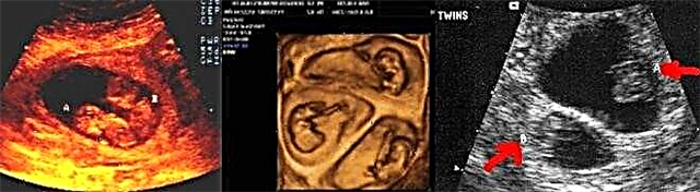
Signs of multiple pregnancy
Usually, it is possible to accurately identify the presence of twins in the uterus only at 8-12 weeks of intrauterine development. In this case, several embryos are well defined in the uterus. They can be located in a wide variety of areas of the uterine space. It depends on where exactly the implantation took place.
It is possible to determine the heartbeat of twins, as a rule, a little later than during pregnancy with one baby. It is possible to establish a heartbeat, but to differentiate how many hearts are beating is a rather difficult task. Usually, the second or third heart becomes audible only by the 20th week of pregnancy. In the early stages, various pathologies are rather difficult to determine in twins.
Is it harmful to the fetus?
There is a huge number of opinions and various myths around the ultrasound scan. Many expectant mothers are worried about the possible harm this study can do to the baby. There is currently no reliable data on the pronounced negative effect of ultrasound on the developing fetus.
Ultrasound screening is performed in many countries. These methods make it possible to identify various pathologies of pregnancy at the earliest possible date. Genetic screening would not have been possible without performing ultrasound diagnostics.
If the expectant mother had cases of chromosomal diseases in kinship, then an ultrasound scan is also a necessity.
If a woman has diseases of the genital organs, then a transvaginal ultrasound scan in early pregnancy can cause her to have a small amount of blood from the genital tract. This condition cannot lead to any complications for the fetus. However, it is worth remembering that if a woman's genital diseases are in the acute stage, then before conducting the study they must be cured without fail.


If the expectant mother has some kind of inflammatory diseases, then transvaginal examination can also lead to a number of complications. Some women have different discharge after ultrasound. Their appearance is possible, mainly, with acute colpitis or vaginitis, which are in the stage of exacerbation.
If a pregnant woman has any unpleasant symptoms in the perineal region, then she must definitely warn her doctor about this before conducting the study.
An ultrasound scan should be performed for all pregnant women at the earliest stages. This not only makes it possible to detect pregnancy in a timely manner, but also to determine the pathological conditions that the expectant mother has.
Often such research is also not worth it. For ultrasound, there are certain regulated deadlines.
For the importance of ultrasound in early pregnancy, see the following video.