Myopia in children (doctors call this disorder myopia) is considered to be a disease of the information technology age. Large amounts of information became available thanks to the Internet, but humanity has paid for it with its eyesight.
Anatomical and physiological causes of myopia in children
- congenital myopia (true, hereditary) - develops either with a sufficiently large anterior-posterior size of the eye (eye length) in relation to the normal or weakened power of the optical system, or with a very pronounced refractive power. In other words, vision with myopia is such that the further away the object is, the more indistinct it seems, since the image is formed not on the retina, but in front of it due to the length of the eye or the structural features of the main refractive media (lens and cornea);
- false myopia in children (accommodation spasm) develops as a result of prolonged contraction of the ciliary muscle, which is responsible for a certain position of the lens, thanks to which it becomes possible to work with objects only at a close distance. Such myopia appears in school-age children due to a violation of the visual regime.
In adults, myopia, manifested at the age of over 40, should also be considered false, since its appearance indicates a change in the density and transparency of the lens substance and is signs of an incipient disease - cataract.
Congenital myopia can already appear in newborns. Whereas the appearance of a false one is characteristic of a school-age child with an increased load on the organ of vision.
Myopia symptoms
The child sees distant objects as indistinct, blurry, blurred, sometimes merging into one point. Some children notice that they begin to squint or pull the upper eyelid more often, as it becomes easier for them to see.

False children's myopia manifests itself gradually. A certain role in the development of refractive error is played by heredity, the period of restructuring of the body in adolescence, ecology, high loads, working with computers and gadgets.
A decrease in distance vision, the appearance of eye fatigue, eye and headaches will make it possible to suspect a child of myopia.
In the case of congenital myopia, it is possible to notice a visual impairment at an early age. The baby does not maintain eye contact with parents and loved ones, bright toys cause weak interest only at close range, the baby does not fix the object and does not try to follow it.
Complications of myopia
Myopia is terrible for its complications, since they somehow lead to a decrease, and sometimes to the disappearance of visual functions.
- Amblyopia... If myopia is uncorrected for a long time, the body does not subsequently perceive the correction, and vision remains low, despite the absence of any organic causes. If one eye sees much better than the other, the worse seeing eye can be turned off from the act of vision, as a result of which strabismus may occur.
- Scleral staphilomas. Due to the increased size of the eyeball, protrusions (staphylomas) can form in places of thinning of the sclera.
- Retinal dystrophic changes. Dystrophic changes in the periphery and in the central zone are caused by an enlarged eyeball and, as a consequence, thinning of the retina at the attachment points and in the central zone. As a result, it is possible to develop hemorrhages, both in the vitreous body and in the retina, as well as a serious complication - retinal detachment.
- Hemorrhages in the vitreous body and retina. It is characterized by the appearance of many black flies, squiggles, nets floating in front of the eye.
- Fuchs spot. This is a pigmented lesion in the area of central vision (macular macula) that arises in severe myopia. His appearance is preceded by hemorrhage in this area. The appearance of a pigmented lesion is most often associated with the formation of the so-called neovascular membrane, which in its structure is a tangle of newly formed vessels. The appearance of a spot is always a bad prognostic sign.
- Retinal disinsertion. Most often, detachment is a consequence of lattice retinal dystrophy. It is characterized by a sudden disappearance of visual functions, the appearance of a "thick curtain", or a veil in front of the eye. Treatment of retinal detachment is only operative.
Methods for diagnosing myopia
Ophthalmological examination begins with determining visual acuity and selecting an adequate tolerable correction. After that, one of the eye drops is instilled, allowing the pupil to expand and block the ability to accommodate. Then the visual acuity is checked again and the correction is selected.
 If a child with myopia is examined for the first time, he will be advised to use eye drops for a certain period, allowing to relieve the spasm of accommodation by forming drug cycloplegia (a condition in which the ciliary muscle stops working and completely relaxes).
After reaching full cycloplegia, visual acuity, correction are again checked and the accommodation reserve is determined. Then refractometry and / or skiascopy are performed. This technique allows you to distinguish true from false myopia.
There are frequent cases when, after carrying out this study, hyperopia was revealed, which was successfully masked under the influence of a spasm of accommodation under myopia.
There are three degrees of myopia:
- weak - 3 diopters or less;
- strong - 6 diopters or more;
- average - from 3.25 to 5.75 diopters.
Determination of the visual field is shown for children with myopia. This study can be carried out for children from 4 to 5 years old. Defects in the field of vision alert the ophthalmologist, because they may indicate complications, one of which is retinal detachment.
All children undergo an examination of the fundus exclusively and only on a wide pupil. The variants of dystrophic changes in the retina determined during the examination are recorded. In case of controversial points or for the purpose of a more detailed examination of certain areas, a Goldman lens is used.
If necessary, for the purpose of visualizing structures, as well as in case of complications, the ultrasound method (B-scan mode) can be used, as well as OCT of the macular region.
Myopia treatment in children
Can a child be cured? There are many techniques and advice on how to treat myopia in children. It should be remembered that the effectiveness of each method depends on the correct selection, taking into account the individual characteristics of the patient.
So, for example, in case of false myopia, it would be strange to start treatment with any surgical methods, and the gymnastics used for the eyes with high myopia, not supported by anything else, will not bring an effect.
Accommodation spasm (false myopia)
False myopia begins to be treated with drugs popularly known as "drops from myopia." The name is not entirely correct, since the drugs used are not an absolute panacea for this refractive error.
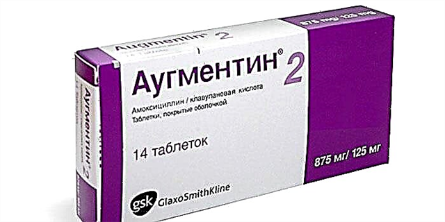
 The eye drops used for myopia are often short-acting mydriatics prescribed by a doctor (Midriacil, Tropicamide, Mezaton, Irifrin), or Cycloplegic (Atropine).
Their main action is to relax the ciliary muscle. Almost all eye drops for myopia are prescription, therefore, their use is controlled by a doctor.
Atropine is a drug that allows you to induce drug cycloplegia and completely relieve the spasm of accommodation.
It is a potent remedy that should be used in doses prescribed by the ophthalmologist.
In order to accelerate the elimination of the drug, it is recommended to drink 1 glass of milk immediately after instilling the eyes. After using this drug, due to the complete relaxation of the ciliary muscle, it becomes impossible to work near, therefore, when carrying out atropinization, the child is given a certificate of release.
The effect of drops can last up to 3 - 5 days after the last instillation.
Short-acting mydriatics (Mezaton, Midriacil, Tropicamide, Irifrin) do not cause partial muscle relaxation.
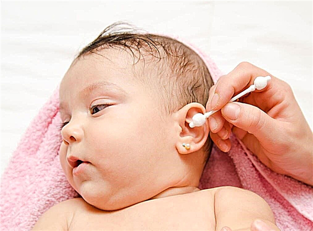
Their use is justified when examining the fundus in children, as well as night eye drops for myopia for children and adults. The short action relieves the ciliary muscle spasm during sleep.
The duration of application of these drops is from 10 to 14 nights in a row, or every other night.
Tear substitutes (Systain-ultra, Optinol, "Artificial Tear", Vidisik) are considered as eye drops to improve vision in case of myopia in cases where a child's prolonged visual stress is combined with work at computer equipment or gadgets.
Moisturizing the eye in a timely manner can relieve fatigue, as well as resist dry eye syndrome.
Exercise for the eyes is used as one of the treatment options, as well as the prevention of myopia in children and adolescents.
True myopia
Almost everyone knows that with myopia in children, correction is used with glasses or lenses. For a long time, it was believed that with myopia, visual acuity should be slightly undercorrected. However, more recently, scientists have proven that it is undercorrection in childhood that leads to the progression of myopia.
 Remember that the use of spectacle or contact correction in case of false myopia with high visual acuity is not recommended.
For the treatment of myopia in children over 5 years old, Paragon orthokeratological lenses are used, which should be worn at night. They allow both to stop the myopia in a child and to cure myopia.
Their action is directed to the cells of the corneal epithelium, due to which the strength of the refractive system changes. Lenses stop disease progression only in cases of mild to moderate myopia. The vision-improving properties are extremely insignificant in severe cases.
Laser surgery techniques (LASEK, LASIK, PRK), which are so popular among adults today, are not recommended for children under the age of 18.
Surgical intervention is aimed at changing the thickness of the cornea, and, consequently, reducing the refractive power in the future. The eyeball grows on average up to the age of 18 years, therefore, surgical treatment at an early age leads to the need for additional correction and additional manipulations.
In some cases, patients over 18 years of age are advised to replace their own lens with a refractive purpose or implant phakic lenses. In the first case, the operation performed is similar to replacing the lens with cataract.
However, a significant difference is the absence of the stage of crushing the lens by ultrasound, since the lens is transparent, and its substance is not compacted.
A silicone lens is implanted into the lens bag, selected taking into account all the patient's characteristics, including both the correction of the astigmatic component and the occupation. In the case of phakic lens implantation of a certain configuration, the lens is placed in the anterior chamber of the eye in front of its own lens.
With a severe degree of myopia, in cases of pronounced degenerative changes in the retina, peripheral laser coagulation of these zones is performed.