From the first days of life, the child gets acquainted with the world around him through sight. Any, even minor, violation in the structure or work of the eye threatens with serious consequences in the future. Compliance with the rules of eye care in young children helps in the prevention of most diseases.
"The child's eyes fester" is a problem that young mothers often face. Under such a serious statement usually lies the presence of an inflammatory disease of the eyelids, conjunctiva or lacrimal system.
What are the symptoms of the disease?
For an eye infection that occurs in infants in the form of blepharitis, conjunctivitis or dacryocystitis, it is characteristic:
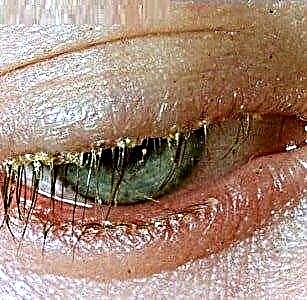
- the formation of a large number of crusts located along the ciliary edge of the eyelids (sour eyes);
- the appearance of purulent contents when pressing on the area of the lacrimal sac;
- redness of the white membrane of the eye (hyperemia);
- the presence or absence of general manifestations of the infectious process, depending on the severity of the disease (increased body temperature, changeable mood of the child, decreased or increased physical activity).
What should be done if an infant's eye festers? You must immediately contact an ophthalmologist. Any delay can negatively affect organ function and lead to disability.
Why does a baby's eyes fester?
In children of the first year of life, the immune system is imperfect, any infectious process can proceed very violently, involving many organs and systems. The slightest non-observance of the rules of personal hygiene can lead to the development of inflammatory reactions.
The baby's eye is festering, what disease can you suspect?
With the exception of eye injuries, there are three main diseases, in the clinical picture of which there is the presence of a purulent process:
- dacryocystitis,
- blepharitis,
- conjunctivitis.
Dacryocystitis
Dacryocystitis is an inflammatory process that occurs in the lacrimal sac, the main cause of which is the obstruction of the nasolacrimal canal.
Due to the development, dacryocystitis is divided into primary (occurring during the first weeks of life) and secondary (developing in older children).
A characteristic difference, with the similarity of the entire clinical picture (conjunctival hyperemia, eyelid edema, purulent discharge, abundance of crusts on the eyelids), is that this disease is one-sided. Almost all mothers note that only one eye festers in a newborn.
 In newborns, the cause of the development of the disease lies in a gelatinous film located in the lower part of the nasolacrimal canal, which should break with the first breath. If this process has not occurred in the baby, then it is the lack of patency of the nasolacrimal canal that is the main reason for the development of the inflammatory process. The child has persistent tearing with the appearance of mucous and purulent discharge with further progression of the disease.
Secondary dacryocystitis occurs in older children. Very often, this disease is a consequence of an untreated dacryocystitis of newborns. The development of this disease is also associated with chronic sinusitis or injuries to the nose or eyes.
Diagnosis of dacryocystitis
In addition to standard diagnostic techniques (determination of visual acuity and field of vision (carried out for children over 4 years old), examination of the fundus, examination in direct and transmitted light)), carry out special procedures:
- Tubular test Vesta. A piece of cotton wool is introduced into the lower nasal passage, while a dye solution is instilled into the eye. A test is considered positive if the tampon stained within 2 minutes. If, after 10 minutes, the paint has not soaked the cotton wool, then the test is considered negative, and the diagnosis of dacryocystitis is confirmed.
- Probing of the nasolacrimal canal in children, for diagnostic purposes, is carried out with great care, since the structures are very delicate, and the reaction of young children is pronounced.
- Lacrimal test Vesta. The lacrimal sac is pre-cleaned by pressing and washing with a 2% boric acid solution. Then protargol solution is instilled. After the child has blinked, the remnants of protargol are cleaned and pressed on the area of the lacrimal sac. With normal function of the nasolacrimal canal, a colored liquid should appear.
- Contrast X-ray of the lacrimal duct allows visualization of the level of blockage in the nasolacrimal canal.
Always, before starting treatment, a smear is taken from the conjunctival cavity for a bacteriological study, which allows you to identify the causative agent of the disease and select antibiotic therapy.
Dacryocystitis treatment
- primary dacryocystitis is easily treated, so if you notice that a newborn's eye is festering, immediately consult an ophthalmologist;
- with a pronounced purulent process, antibiotics are prescribed in the form of drops, taking into account the sensitivity to them;
- The main method of treatment is considered to be the massage of the lacrimal sac area, the technique of which is taught by an ophthalmologist. Massage should be performed 5 times a day, before feeding the baby, for 2 weeks. Do not try to massage before consulting your doctor. Remember that one wrong move can do more harm than good;
- in cases where the desired effect has not been achieved, you will be advised to rinse the lacrimal ducts;
- if the patency could not be restored, then the next step will be to perform sounding, or bougie of the lacrimal canal to eliminate the obstacle and ensure lacrimation;
- in severe cases, surgical treatment is indicated - dacryocystorhinostomy, the conduct of which is aimed at creating an artificial nasolacrimal canal;
- secondary dacryocystitis is treated only promptly.
Blepharitis
 blepharitis is an infectious-allergic disease, manifested by an inflammatory process in the region of the eyelid margins;
- often an abundance of crusts, sticking of eyelashes and edema in the area of inflammation - create a picture of suppuration, or suppuration of the eye;
- the main cause of the onset of the disease in infants is a violation of the rules of personal hygiene.
 blepharitis is an infectious-allergic disease, manifested by an inflammatory process in the region of the eyelid margins;
Blepharitis, depending on the form of the disease, is divided into scaly, ulcerative, angular, meibomian and demodectic.
- Ulcerative blepharitis tends to develop mainly in children. It is characterized by severe pain syndrome, since under the scales on the eyelids there are always sores that bleed.
- In adolescents, angular blepharitis is most common, a feature of which is the presence of foamy contents, scales and ulcers in the corners of the eyes.
- Demodectic blepharitis, caused by a tick of the genus Demodex, occurs with the same frequency in both adults and children and is manifested by profuse loss of eyelashes, the follicles of which the tick feeds on.
Diagnosis of blepharitis
Before treatment, the necessary examination is carried out:
- standard diagnostic techniques: determination of visual acuity and field of vision (carried out for children over 4 years old), examination of the fundus, examination in direct and transmitted light));
- special diagnostic examination - microscopy of crusts and eyelashes.
Always, before starting treatment, a smear is taken from the conjunctival cavity for a bacteriological study, which allows you to identify the causative agent of the disease and select antibiotic therapy.
Treatment
- if the eye festers or the eyelid suppuration occurs, then antibacterial ointments are prescribed, which must be applied at least 4 times a day;
- to improve the regenerative properties of the skin, the edges of the eyelids are lubricated with sea buckthorn oil;
- smearing the edges of the eyelids with a solution of brilliant green is not recommended for children because of the high probability of developing a chemical burn of both the eyelid and the eye itself;
- with demodectic blepharitis, special ointments (zinc-ichthyol, metronidazole, and others) and gels for washing are prescribed, aimed at eliminating the tick. They are used continuously for 25 days, after which they re-examine the eyelashes and scales.
Remember that self-medication can be dangerous to your health, be sure to consult an ophthalmologist.
Conjunctivitis
- conjunctivitis is a disease in which the inflammatory process occurs in the conjunctiva;
- the immune system in children is prone to hyperreactive responses to any infectious agent, therefore, even in newborns, the eyes can fester;
- in children, untreated conjunctivitis can lead to complications;
- if the baby's eye festers, then his general condition worsens: the child is capricious and tries to rub his eyes;
- despite the fact that purulent discharge is characteristic of bacterial conjunctivitis, the course of any conjunctivitis may be complicated by the addition of an infectious component;
- usually epidemic conjunctivitis is common in autumn and spring, but it can also occur in cold rainy summers. Conjunctivitis can be infected by contact - through personal belongings.

The disease is most severe at an early age. In addition to the fact that the baby's eyes fester, there is often a general response of the body: chills with a sharp rise in temperature to high numbers, lethargy, adynamism.
- the eyes of an infant can fester in the presence of an inflammatory process in other organs and tissues. In this case, the development of conjunctivitis is considered as a manifestation of a septic reaction, prescribing massive antibiotic therapy;
- initially one eye is pounded, the process becomes two-sided after 2 - 3 days. The discharge is purulent, its color can vary from yellow to green, the presence of many crusts along the ciliary edge of the eyelids, pronounced blepharospasm is characteristic. Epidemic conjunctivitis is always differentiated from diphtheria, a feature of which is the presence of practically non-removable crusts along the edge of the eyelids and membranes on the conjunctiva. If you still try to remove them, the underlying tissues begin to bleed heavily;
- a special group of conjunctivitis caused by sexually transmitted infections - gonococcal and chlamydial - are another reason due to which the eyes of a newborn fester.
The baby is infected at the time of delivery. Development is fast, lightning fast. Serous discharge during the day becomes hemorrhagic, and then purulent with a pronounced green color.
A characteristic feature is the bleeding of the conjunctiva upon contact with it. A corneal ulcer almost always develops, characterized by a high probability of perforation with subsequent death of the eye. Visual functions are not restored.
Diagnosis of conjunctivitis
Standard diagnostic techniques are used (determination of visual acuity and field of vision (carried out for children over 4 years old), examination of the fundus, examination in direct and transmitted light).
Conjunctivitis treatment
What to do if an eye is festering in a baby or newborn?
- always, before starting treatment, a smear is taken from the conjunctival cavity for a bacteriological study, which allows to establish the causative agent of the disease and select antibiotic therapy;
- conjunctivitis is not treated on its own, even with a relatively mild course, consultation with an ophthalmologist is necessary. The self-prescription of antibacterial and anti-inflammatory drops threatens to help with the development of complications. Traditional medicine recipes very often lead to undesirable consequences, up to and including loss of the organ of vision;
- drug treatment is based on the use of antibacterial eye drops. Their main active ingredients are fluoroquinolones (recommended for use from the age of 7) or aminoglycosides (used from birth). However, with a pronounced infectious process, when the risk of losing an organ of vision is higher than possible side reactions, antibacterial drugs can be used regardless of the child's age. Eyes are often buried - up to 8 times a day.
Prevention of conjunctivitis in newborns in the hospital
Prevention of chlamydial and gonococcal conjunctivitis is carried out by prescribing prenatal sanitation for pregnant women, followed by treatment and instillation of antiseptics and antibacterial drops into the eyes of newborns immediately after birth.

It should be remembered that even with the classic picture of the inflammatory process, one should not discount the injury to the organ of vision. The risk of infection of internal structures is high. In this case, timely prescribed treatment is the secret of a successful cure!
Proper child care, early training in personal hygiene rules at two or three years, tempering, stimulating immunity will protect you and your child from such a formidable group of infectious eye diseases, which will preserve vision for many years!