Assessment of the lengths of bony landmarks is a necessary diagnostic procedure performed by a specialist for all women while carrying a baby.
The boundaries and structural features of the pelvic apparatus have been evaluated in expectant mothers for many centuries. Such a simple and informative study allows doctors to obtain a lot of the diagnostic information they need.
A little about anatomy
The pelvis is a bone formation. Quite a lot of different bones and joints are involved in its formation. The pelvic bone apparatus is a complex architectural element. Each woman has her own characteristics of his anatomy.
The pelvic bone apparatus is formed by several bones at once: a pair of pelvic, sacral and coccygeal. Each pelvic bone, in turn, consists of three more: the iliac, ischial, and pubic. They are interconnected by cartilage tissue.
This arrangement is functionally beneficial during pregnancy. It helps the baby to move calmly along the birth canal.

The pelvis is a kind of receptacle for the reproductive organs. During gestation and childbirth, it has a very important function. It is in it that the birth canal passes, along which the baby subsequently moves during his birth into the world.
Determining the size of this bone apparatus is very important. It is especially important to do this if the baby is not physiologically located in the mother's womb. Breech presentation of a child with a narrow or asymmetric mother's pelvis requires a more attentive attitude to the woman during the course of her pregnancy.


Determination of clinical parameters
For many years, doctors have performed external examination of the pelvis in different ways. The first of them is the determination of the indicators of the pelvis by palpation. The second method consists in determining the studied lengths using a special device - a tazometer.
Doctors carry out this diagnostic procedure when carrying a baby. at least twice... For the first time, these clinical indicators are determined at the very beginning of pregnancy. The obtained values must be included in the personal medical card of a pregnant woman. Usually, the measurement of the pelvis is carried out for women who are registered for pregnancy.


Also, doctors determine the size of the pelvic bone apparatus in expectant mothers already at a period closer to childbirth. This is a very important prognostic indicator for assessing how the labor will go. He also helps doctors choose the best method of obstetrics needed for a particular patient.
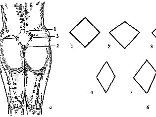
When conducting a study, the doctor will be especially interested in a special anatomical zone - Michaelis rhombus. This site is located in the lumbosacral segment of the spine.
Its changes are a very important diagnostic criterion for doctors.


The obstetrician-gynecologist measures the size of the pelvis, who will further observe the woman for 9 months of bearing her baby. The research is carried out in a regular office.
Measurement of the pelvis is performed while the expectant mother is lying on the couch. The starting position of a pregnant woman is on her back. In order to simplify the diagnostic procedure, the expectant mother should lift the clothes from the measured area. To determine the indicators, the doctor uses a pelvis meter.

How is the norm determined?
An obstetrician-gynecologist measures several sizes at once. One of them is longitudinal. And the other three are transverse. Each of these values has its own standard criteria. They are used by doctors in order to accurately determine the type of structure of the pelvic apparatus in a particular patient.
Several of the investigated parameters are called by a special term - Distantia or D. To determine the first of them, doctors measure the distance between both trochanteric zones of the thighs. They call this parameter D. trochanterica. For most women, its values are in the range from 28 to 33 cm.
To determine the next investigated parameter, the distance between the scallops of the iliac ossicles is determined. It is called D. cristarum... Its normal values are in the range from 24 to 27 cm.


Another equally important determinant is the external conjugate. To determine it, doctors measure the distance from the upper part of the bosom to the edge of the final lumbar region (at the level of the fifth vertebra). Its values range from 20 to 21 cm.
After the measurement, the doctor can calculate true conjugate. This indicator is less than the outer one by 9 cm.
In medical practice, there is another method for determining this size. For this, the doctor must determine the diagonal measurement. For this purpose, he measures the distance between the most protruding point of the sacral promontory to the lower edge of the symphysis.
More often, this clinical indicator is determined during palpation by a gynecologist on a chair. Its norm is 10-13 cm.


The physician can still measure the direct measurement of the pelvic outlet. For this, the distance from the apex of the coccygeal bone to the lower angle of the bosom is measured. This indicator is equal to eleven centimeters.
To refine this parameter, another refined criterion is also used - true straight measurement. Its norm is already nine and a half centimeters. The mathematical difference between these two determinable dimensions is, as a rule, one and a half centimeters.

Angle of inclination of the pelvis is also a very important clinical indicator. Two planes are involved in its formation horizontally and vertically. In order to determine this clinical criterion, a tazouglometer is used. In the vertical position, the normal values of this determined parameter are 45-50 degrees.
During the study, the doctor can also additionally determine several other sizes. They have additional diagnostic value. Usually they are necessary in order to identify the individual features of the structure of the bone apparatus, which are available in a particular patient.


If, when determining the size of the pelvis, the specialist has determined any asymmetry, then he will also additionally measure the following parameters. They are presented in the table below:
Clinical options
The doctor takes into account the ratio of all these indicators. This allows him to assess the type of pelvis in a pregnant woman. For this, several sizes are assessed at once: a specialist does not make a conclusion based on only one clinical parameter.

The table below shows the different types of pelvic structures in women:
How is the decoding of the obtained values carried out?
If the pelvis has a normal structure, then the Michaelis rhombus looks like a square that is inverted. Its diagonal is about 11 cm.
When measuring this indicator, it happens that the sides of the square begin to shift. This also leads to a change in its shape: it becomes more elongated. If, during the measurement, the doctor determines a pair of acute and a pair of obtuse angles, then in this case it means the presence of a narrow pelvic bone apparatus.
A wide pelvis is most often found in fairly tall and large women. This is influenced by the structural feature of the musculoskeletal system of the expectant mother. Also, a wide pelvis can also be found in women with an average physique. In miniature ladies and expectant mothers with small stature, such a structure is practically not found.

A wide pelvis is characterized by an increase in all determined sizes. When measuring dimensions, it is very important to exclude the influence of a large amount of subcutaneous fat. For this exception, a gynecological examination is performed on the chair. By determining the true conjugate, the physician can determine how wide the pelvis is in a particular patient.
Many expectant mothers think that the larger and wider the pelvic bones, the easier it will be for them to give birth on their own. This is not entirely true.
Indeed, the size of the pelvic bone apparatus is of great importance for the possibility of natural childbirth. However, in the case of a wide pelvis, the expectant mother may experience various pathologies.

Also this is no exception. for the appointment of a caesarean section. Surgical delivery can be indicated with a capacious and deep structure of the pelvic apparatus. The choice of the method of giving birth is determined by the obstetrician-gynecologist who monitors the course of pregnancy.
Symmetry - this is a very important parameter that the doctor must record. There is a certain medical algorithm for this. The doctor should measure the measurements on both halves of the torso. If the obtained size values on the left side are larger than the right-sided ones by 1 cm or more, then in this case the doctor records the presence of asymmetry.
It is also important to evaluate the measured side dimensions. To do this, the doctor will measure the distance between the edge of the anterior superior and posterior superior bones. These clinical parameters are determined on both the left and right sides. Normal values for this indicator are 14 cm.

If the obtained values are significantly less than 12.5 cm or are clearly different from each other, then this also indicates the presence of asymmetry in the pelvis of a pregnant woman. In such a situation, the bones are displaced in the vertical plane.
Doctors also call this variant of the structure of the pelvic apparatus asymmetric. In this situation, a caesarean section is usually required. Natural childbirth can be dangerous for both the woman and her baby. The risk of various injuries in this case increases many times.

How to measure yourself at home?
You can try to measure the size of the pelvis without the participation of a doctor. However, such measurements can only be indicative. Still, the type of structure of the pelvis and its main dimensions are determined by an obstetrician-gynecologist who observes the course of pregnancy in a particular woman.
The specialist has the necessary experience and knowledge to successfully carry out this important diagnostic procedure.

It often happens that the expectant mother wants to independently determine what kind of pelvis she has. To do this, she simply measures the circumference of the thighs or the distance between the most distant bony formations of the pelvis.
This measurement has nothing to do with the clinical determination of the size of the pelvic structure. A comprehensive and full-fledged study can only be carried out with the participation of a doctor.

For information on how to measure the size of the pelvis during pregnancy, see the next video.