Informativeness of the MRI procedure in children
The information content of the method depends on the tissue that is being examined. Magnetic resonance imaging better than CT "sees" soft tissues and accumulations of fluids in cavities. This procedure is absolutely safe for children.
Why the popularity of MRI is increasing every year
The magnetic resonance method began to be used in medicine since 1946.
It has become widespread, since it has a number of positive aspects:
- non invasive procedure, which makes it possible to use this method. Including in pediatric practice and neonatology;
- possibility getting a picture from any part of the body a person;
- no radiation exposure.
On the finished image, you can determine malignant neoplasms, fluid accumulations in tissues or cavities, the integrity of organs, etc.
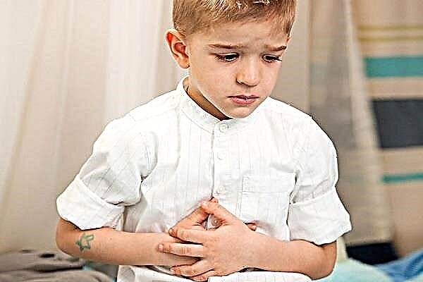
Today, MRI for children is widely prescribed for examining the peripheral and central nervous system (for example, to clarify the diagnosis for headaches of unknown etiology, other neurological symptoms), the musculoskeletal system.

Method essence
The method is based on the phenomenon of magnetic resonance. The magnetic properties of the nuclei of some elements allow them to reorient themselves in a magnetic field in a certain direction, and this direction is always constant. Under the influence of a magnetic field, atoms tend to move to a lower, more favorable energy level.
The stronger the magnetic field, the more the core is polarized. If some energy is imparted to such a system, then the system will leave an energetically favorable state, for some time it will be in a nonequilibrium state, but under the influence of a magnetic field it will return to its initial energetically favorable state with the release of energy.
Since different substances must be given different energies to get out of the equilibrium state, substances can be distinguished by the magnitude of this energy. This energy is converted into a signal, and the computer builds an image based on the received data.
Who is MRI assigned to?
MRI can be prescribed by a wide range of specialists: therapists, surgeons, oncologists, gastroenterologists, cardiologists, etc. adults and children of any age and gender. The direction for MRI for children is written, respectively, by the pediatrician.
In each case, the doctor determines the individual indications for the study for the patient.
Contraindications for performing in children

Contraindications for MRI studies are general:
- the presence of metal prostheses, teeth, screws, plates, brackets, piercings and other metal structures in the human body;
- fear of confined space;
- allergy to medications used.
Does the child need special training before performing an MRI
Special preparation is required only for the examination of the abdominal cavity organs (diet excluding foods that stimulate gas formation in the intestines) and MRI of the pelvic organs (the bladder must be full).
Other localizations do not require preparation.
MRI under general anesthesia. Is there a recovery period after the procedure
For children, MRI under anesthesia is indicated if the child is panicky about the upcoming procedure or if he has diseases of the nervous system, in which it is not possible to be immobile for a long time. Also, an indication for general anesthesia may be mental illness of the subject. And, of course, age - very young children simply cannot be in one position for a long time.
As with usual surgical anesthesia, it is impossible to perform activities requiring attention and concentration in the next 24 hours after the procedure.
If the study is carried out under anesthesia, special training is required. The last time the child can be fed 6-8 hours (children up to a year for 4 hours) before the study. No need to give in to the child's requests and feel sorry for him! Even the smallest candy consumed before anesthesia or drunk juice can lead to the development of fatal complications during anesthesia!
What allows you to detect MRI, or Read the results
MRI allows you to identify structural changes in organs and tissues of the body, congenital and acquired malformations.
Brain and neck
MRI of the brain allows you to identify foci of abnormal nervous tissue (tumor, hemorrhage, ischemia), aneurysms, signs of increased intracranial pressure, to assess the condition of the vessels of the head and neck, lymph nodes and neck organs.
Rib cage
It is possible to diagnose and assess the condition of the lungs, trachea, heart with surrounding tissue, esophagus, aorta, etc. An assessment of the prevalence of tumors of the chest organs, an assessment of the stage of development of inflammatory processes, the state of lymph nodes and blood vessels are available.
Abdominal cavity and pelvic organs
MRI of the pelvic organs can help in the differential diagnosis of gynecological diseases and surgical ones. Using the method, you can determine ectopic pregnancy, establish fetal malformations, determine the presence of pathological formations (tumors, cysts) in the uterus and ovaries, etc.
MRI of the abdomen can be done with contrast enhancement. This study is informative for therapists, gastroenterologists, surgeons and oncologists. This study significantly expands the possibilities of diagnostics and differential diagnostics of diseases of the abdominal organs.
Limbs
MRI of the extremities will show pathological processes in the joints, bone tissue. Using this method, it is possible to identify fractures, damage to bones, ligamentous apparatus, and neoplasms of bone tissue.

Where can an MRI be performed and what conditions are needed for this?
Referral from a specialist
The referral of a specialist is filled in according to form 057 / y. The form contains the last name, first name, patronymic of the citizen, year of birth, place of registration and residence, insurance policy number and insurance company, diagnosis code according to ICD-10. The referral is substantiated by a doctor, certified by signatures and seals.
Even paid research is possible only with a referral!
MRI free
Free MRI can be done only in public hospitals with a doctor's referral. The study is expensive due to the high cost of equipment and high energy costs required for the procedure. Private clinics carry out this research only on a commercial basis.
For MRI, you only need a passport, policy, SNILS and a doctor's referral for free.
It is possible to carry out this study on the basis of compulsory medical insurance, but you may encounter some difficulties. This study has a limited range of indications and the attending physician must clearly justify the patient's referral for tomography. After receiving a referral, the patient is placed on a quota queue. The queue can only be moved by the head physician of a medical institution for a number of indications.
The second difficulty is that not every compulsory health insurance policy can provide this procedure free of charge. If the insurance company has this type of research on its list, then the patient can apply for a free service.
If the insurance company has not included in the list of free MRI examinations, then, alas, the research is carried out only in a paid clinic or in a government institution with an appropriate license that allows you to provide paid services.
Payable service. Price depending on the area of study
The cost of the study depends on the clinic in which the study is conducted, the complexity and duration of the study, the study area, whether contrast will be used during the study, and many other factors.
Approximate average values can be cited as an example (since they vary greatly from city to city).
For example, an examination of the head and neck, spine can cost in the range of 3-5 thousand rubles; MRI of soft tissues - about 4 thousand, MRI of the abdominal cavity and retroperitoneal space - up to 8 thousand, etc. In addition to the cost of the service, the image recording to disk, the issuance of duplicates, etc.
MRI for pregnant women

Conducting MRI for pregnant women is not contraindicated; on the contrary, the method improves the quality of prenatal screening (in addition to ultrasound methods), and reduces the number of invasive interventions. Research results, detection of malformations, for example, can help in determining the tactics of treating a newborn.
As for the diagnosis of maternal diseases, the study is permissible if indicated, since it is safe for the fetus.
MRI can help in the diagnosis of ectopic pregnancy, predict the course of pathological pregnancy in the presence of fetal malformations.
Conclusion
Non-invasive research methods are widely used both in outpatient care and in hospitals, no matter whether adults or children. An MRI scan can be done for a child free of charge or for a fee, subject to the appropriate indications and a referral from a doctor.
The method is expensive (the high price of the equipment, expensive maintenance and the power consumption of the device is large), but in difficult cases it is necessary to clarify the diagnosis in addition to clinical data and physical examination of the patient.
Large centers have more opportunities to use this method; specialists located in the periphery, sometimes it is not possible to prescribe this method for their patient. Many organizational tasks still need to be addressed.