What is epidermolysis bullosa?
Epidermolysis bullosa includes a huge number of skin diseases that are inherited and have a common feature - blisters of different sizes and different numbers. They are formed on the skin or mucous membranes due to any, sometimes minimal impact. Any friction, touch, change in temperature and air humidity can aggravate the process.
The disease can manifest itself at different ages. The child becomes ill while still in the womb. Most often, he is either already born with signs of the disease, or they appear soon after birth. In all cases, it is more correct to call the disease congenital epidermolysis bullosa.
This is a genetic disease, that is, it is not transmitted from a sick person under any circumstances and in any way. The baby can only inherit it from mom and dad at the time of conception.
How many people have epidermolysis bullosa?
Most often, the disease is recorded in children aged 1 to 5 years. Data from the National Epidermolysis Bullosa Registry, which is conducted in the United States of America, suggests that 1 in 50,000 newborns suffers from the disease. Over the 16 years of its existence in the United States, 3300 people have been identified with this disease.
In Europe, epidermolysis bullosa affects 1 in 30,000 newborn babies. Japan has the lowest prevalence of the disease, it occurs in 7.8 out of 1 million children born.
Unfortunately, there are no official statistics on the disease in Russia. It is known that in our country, Ukraine, Belarus and Kazakhstan, there are more than 150 patients with epidermolysis bullosa.
Causes of epidermolysis bullosa
The cause of this serious illness is mutations in genes that are responsible for the synthesis of structural proteins in the skin. As a result, its cells lose strong connections with each other. The slightest impact from the outside contributes to damage to the skin.
Human skin consists of several layers. Upper - the epidermis is represented by keratinocyte cells. They constantly divide and, as they grow, move from the underlying layers to the upper stratum corneum, providing renewal of the skin and its protection. Keratinocytes are connected to each other by special bridges - desmosomes, from which protein filaments - tonofibrils - protrude. The cells of the lower layers of the epidermis are also connected by the protein laminin.
The epidermis is followed by a layer called the dermis. It includes collagen, elastic and reticular fibers, which are permeated by numerous blood and lymph vessels, nerves, sweat and sebaceous glands, hair follicles. The dermis contains fibroblast cells. It is they who produce all the fibers in the layer.
The dermis and epidermis are firmly connected by a layer - the basement membrane. It also contains collagen proteins. They are the main components of the retention threads. These filaments tightly attach the basement membrane with the covering epidermis to the dermis.
Various clinical forms of epidermolysis bullosa are based on structural disorders of the skin, when, as a result of mutation, the growth and maturation of damaged keratinocytes occurs, the absence or decrease of retaining and fastening protein threads. A deficiency of certain proteins involved in the structure of the skin is also possible: collagens, keratins, laminin and others.
As a result, the connections between the layers of the skin and its cells are weakened, and at the slightest external influence, damage occurs with the formation of bubbles.
Epidermolysis bullosa classification
Since the development of technologies that make it possible to determine the structure of the skin at microscopic levels and determine its smallest structures, epidermolysis bullosa has been divided into 3 groups. Later, another group of the disease was identified.
In the modern medical community, the classification of the disease consists of 4 main groups and 6 subgroups. In subgroups, various clinical forms of the disease are collected. They differ in the type of inheritance, microscopic changes in the skin, clinical manifestations, severity, prognosis.
So, epidermolysis bullosa can be simple, borderline, dystrophic. Kindler syndrome is a separate group. Simple epidermolysis bullosa includes suprabasal and basal. The forms of borderline epidermolysis bullosa can be localized and generalized. Dystrophic epidermolysis bullosa is dominant and recurrent. Kindler syndrome is not divided into subgroups.
The classification takes into account the layer of the skin where the bladder forms, as well as changes in the proteins that make up it.
Epidermolysis bullosa manifestations
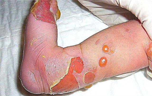
Blisters, erosions of various sizes on the skin and mucous membranes are the main sign of epidermolysis bullosa. They appear due to a decrease in the skin's resistance to various influences from the external environment. This often happens when the ambient temperature changes, the effect of pressure and friction. Due to the fact that the skin has an unusual structure, bubbles appear, then erosion. Their healing with some types of epidermolysis bullosa can take place with the formation of rough scars.
Other manifestations of epidermolysis bullosa often include changes in the color of the skin, keratosis on the palms and soles, contractures of the small joints of the hands, and fusion of fingers. Less common are baldness, or partial hair loss, increased perspiration, or, conversely, its absence, tooth damage, difficulty swallowing, vomiting, constipation, diarrhea.
Simple epidermolysis bullosa
Babies with simple epidermolysis bullosa are born with blisters on the skin, or they appear in the first months of their life. Blisters can be seen on the hands, feet, elbows, knees, legs, scalp. In the oral cavity there are few or none at all. Blisters and erosions are painless and heal quickly.
The nails do not change. If their detachment occurs, they must be restored. The teeth of such children are healthy. The blisters heal without leaving a trace. As the child grows, they will form less and less.

Localized epidermolysis bullosa simplex of the hands and feet refers to basal epidermolysis bullosa simplex. It manifests itself with the beginning of independent walking. The disease can also begin in adolescents, when wearing tight shoes begins and the feet are injured. They are most often affected. The defeat of other parts of the body is less common. Their number is different, from minor rashes to huge, limiting ordinary life.
Generalized epidermolysis bullosa is also one of the clinical forms of basal epidermolysis bullosa. In babies, the back of the head, back, elbows are affected. As the child grows and matures, bubbles appear on the hands, feet, and places subject to friction.
There are many bubbles. They are arranged in groups that form centers of various bizarre outlines. Mucous membranes are affected, nails come off easily. Skin pigmentation often changes, hyperhidrosis and hyperkeratosis of the palms and soles appear. Important - no scars remain after skin damage.
Usually, when the ambient temperature rises, the condition of patients with epidermolysis bullosa worsens. However, in babies with a generalized form of epidermolysis bullosa, an increase in temperature has a positive effect on the condition of the skin.
Borderline epidermolysis bullosa
With borderline epidermolysis bullosa, changes affect the basement membrane of the epidermis. It is subdivided into generalized and localized. A common feature is the appearance of blisters on any part of the body with extensive lesions. Changes in the tooth enamel are also characteristic, it becomes thinner, point depressions appear on the surface of the tooth. They are often exposed to tooth decay. Borderline epidermolysis bullosa is more severe than simple.
Generalized severe borderline epidermolysis bullosa is also called "lethal". This is a fatal disease, the outcomes of which are gross disfigurement and disability. The baby is born with bubbles, or the age of their manifestation is the neonatal period. Blisters are often localized in the perioral region.
They cover the scalp, legs, perineum, chest of the child. They rarely appear on the hands and feet, unlike other types of epidermolysis bullosa. The exception is the terminal phalanges of the fingers, on which the nails are located. The plates themselves collapse, peel off and are lost forever. All mucous membranes are often affected by the rash.
Erosions heal very slowly. In their place, the skin atrophies, rough scars form. Due to the complications that have arisen, the growth of babies stops. They don't gain weight. Such children often die before even three years from infections, exhaustion and circulatory disorders.
Generalized moderate - severe borderline epidermolysis bullosa differs from the previous variant in a milder course. Bubbles can also be found in a newborn baby, but no scars form when they heal. A distinctive feature of this clinical variant is focal hair loss and severe atrophy of the scalp. Such babies grow and develop in accordance with age. The disease does not affect this in any way.
Dystrophic epidermolysis bullosa
This group of epidermolysis bullosa is classified into two subgroups depending on the type of inheritance: autosomal dominant or autosomal recessive. This means that the genetic mutation is clearly inherited. In the first case, for the development of the disease, only one mutant gene is sufficient, which is in the father or mother of the child. In the second case, both parents must be carriers of the defective gene, but they themselves will be healthy. But the baby will be born with a serious illness.
Dominant dystrophic epidermolysis bullosa is characterized by the formation of blisters on all parts of the body and mucous membranes from birth. In some clinical cases, blisters develop even later. They are prone to recurrence but heal quickly with scarring and hypopigmentation. This form of epidermolysis bullosa does not affect the growth and development of the child.
Recessive dystrophic epidermolysis bullosa more often than other forms of the disease leads to severe disability, although its severity may not be so severe. Blisters can be located on the hands, feet, elbows, and knees, or spread throughout the body. Similar changes appear on the mucous membranes. Due to the formation of rough scars, the baby becomes disabled.
Kindler syndrome
Children with Kindler syndrome are born with blisters on the skin and mucous membranes. At an older age, photosensitivity appears, pigmentation on the skin, scar tissue forms, and nails peel off. Characterized by damage to the gastrointestinal tract and urinary tract.
Complications of epidermolysis bullosa
The development of complications often develops in children suffering from borderline and dystrophic epidermolysis bullosa. Diseases of these groups occur in the most aggressive forms.
Extensive ulcers and erosions on the skin over time lead to the fact that it is replaced by rough scar tissue. It is characterized by poor sensitivity, weak extensibility, lack of sebaceous and sweat glands. The functions of the skin are lost, in addition, scars are also a cosmetic defect that disfigures the appearance.

Cicatricial replacement of the eyelid skin leads to limitation of their movement. The kid will not be able to fully open or close the eye, as a result of which the organ of vision may be left without protection. The shape of the eye will also change until it is completely closed. As a result, conjunctivitis, blepharitis and other inflammatory diseases of the eye join. The child may lose sight altogether.
Scars in the mouth lead to the formation of a microstomy. The mouth slit is pulled together, narrowed. As a result, the baby will not be able to open his mouth to the end. The process of swallowing and speech is impaired.
Coarse scar tissue in the area of the joints leads to limited movement in them. The child will not be able to fully bend and straighten the joint, since the scar will not be able to fully stretch like normal skin. The joint often remains in the same position, its contracture develops - stiffness.
Erosion and wetness in the area of the fingers lead to the fact that scar tissue forms with the fusion of the fingers. In this case, the child's hands will forever lose their grip function.
Erosions on the mucous membranes can also heal with the formation of scar tissue, leading to narrowing - strictures of the esophagus, respiratory and urinary tract, intestines. The child cannot fully swallow food, speak, breathe, urinate, therefore, he will lose weight up to cachexia, often get sick with inflammatory diseases of the lungs and kidneys. The absorption of food in the intestines will be impaired. This will further aggravate the baby's condition.
Ulcers and erosions on the skin are the gateway to various infectious agents. Therefore, if the dressing rules are not followed, purulent inflammation of the skin may occur, and sometimes systemic blood poisoning - sepsis.
People with epidermolysis bullosa are at high risk of developing squamous cell skin cancer.
Life expectancy with epidermolysis bullosa
The lifespan of butterfly babies depends on the form of the disease. The most favorable prognosis for babies suffering from simple epidermolysis bullosa. They can even fully live and learn. There is an option that with age, the disease will generally recede.
Generalized borderline epidermolysis bullosa is characterized by the most severe course. Children with this form of the disease sometimes do not live up to three years old due to the development of complications incompatible with life.
In other forms of the disease, in most cases, severe disability develops. But babies still have a chance to live a long life with proper supervision and care.
Diagnosis of epidermolysis bullosa
The diagnosis of epidermolysis bullosa cannot always be made only on the basis of complaints. To confirm or refute it, complex laboratory diagnostics are needed in large clinics. Genetic analysis is also required.
A skin biopsy is mandatory, always from a fresh bladder. A piece of skin should be immediately frozen in liquid nitrogen, or soaked in saline. For long-term storage, a piece of skin is placed in a special transport medium. The biosample is examined under special powerful microscopes with contrast agent processing. It is possible to determine abnormalities in the structure of the skin, and to identify the lack of certain protein components.
Before the baby is born, genetic analysis plays an important role. It does not affect treatment, but it is needed by the family where a child with epidermolysis bullosa is already being raised, and where a second pregnancy is planned.
Treatment principles for epidermolysis bullosa
In the modern world, epidermolysis bullosa is considered an incurable disease. Treatment and prevention actions should prevent trauma to the skin.For this, the skin of "butterfly children" needs the right care. For the comfort of young patients, it is important to eliminate itching and pain.
To prevent life-threatening complications, it is necessary to deal with skin infections in a timely manner. If complications develop from the digestive system and joints, you need to correct them in time. Parents of butterfly babies should be trained to care for them as soon as possible. For him, special soft plasters, bandages, ointments with silver, antiseptics are used.

Large bubbles usually burst gently. The necessary agent is applied to the resulting erosion. Then the wound is closed with a bandage. They use specialized atraumatic bandages that contain a sorbent, an antiseptic, regenerating and antimicrobial agents.
Special hydrogel and hydrocolloid films have also been developed. They cover erosion and ulcers, thereby preventing drying out. You can use porous collagen sponges. They can stick tightly to the wound, and separate on their own if it gets very wet or heals completely.
A second bandage is applied on top. Thanks to her, the medical bandage will be well fixed and will sit tightly in place. The fixation bandage should not press hard on the skin and be tied to a tight knot so as not to injure the skin even more. To prevent complications, separate dressings for each finger of the child are needed. The limb when bandaging should not be bent or unbent. Dressing a baby with severe clinical variants of epidermolysis bullosa can take 1 - 2 hours.
A baby suffering from epidermolysis bullosa should be observed by many specialists and regularly examined by a dermatologist, pediatrician, gastroenterologist, otolaryngologist, surgeon. If necessary, consultations of a thoracic and plastic surgeon are needed.
In some cases, an antibiotic should be administered in injections. The child should receive all the necessary nutrients and vitamins with food. Children are assigned specialized high-protein mixtures. If, due to complications, the baby cannot swallow food normally, a gastrostomy may be installed.
To help mothers and fathers of special babies, a “butterfly children” fund has been created. He helps in the treatment and rehabilitation of children suffering from epidermolysis bullosa, buys medicines for them. If psychological and legal assistance is needed, it will also be provided thanks to the fund. Its specialists also help doctors who are faced with illness.
Conclusion
Epidermolysis bullosa is a serious and, unfortunately, incurable disease. All treatment activities are palliative only. But with the correct and timely started therapy, you can significantly alleviate the suffering of the baby and give him a chance for a long life.